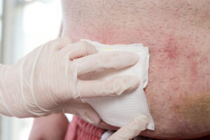
The term debridement takes its origin from the French language and dates back to 1839. The word debridement literally means “unbridling” or to unbridle. The term bridle refers to a horse’s harness used to restrain or govern the animal. The prefix de- is also inherited from the French language and functions in reversing a verbs action and hence, the removal of the bridle. In present day vernacular, debridement is referred to as the removal of dead, damaged, devitalized, contaminated, or infected tissue. But what is the purpose of debridement and how is it performed?
Our first step in wound debridement methodology is to establish what type of wound we are dealing with and look at the etiology or the root causal factor. For instance, in patients with diabetes we often encounter diabetic foot ulcers located on the plantar surface of the feet which are primarily due to a combination of repetitive pressure in the presence of peripheral neuropathy or lack of a pain threshold. This type of wound usually and often presents with thick hyperkeratosis as evidence based on pressure and occurs in areas of primary weight bearing such as the sub-metatarsal platform, heel, and even the digital areas especially the hallux. We encounter venous stasis ulcerations predominantly in the lower extremities due to excessive swelling where the ulcer is usually broad, superficial, weeping and extremely sensitive. These ulcers commonly occur in and around the medial and lateral malleoli due to the lack of supportive tissue in conjunction with a conglomeration of large and superficial venous structures. We see yet a third type of wound in areas of the lower extremities secondary to a lack of blood flow (vascular ulcerations) that present with a punched out border with an escharotic base associated with cold and rigid epidermal texture and turgor, and the list goes on. In essence, the clinician must not only have to know the methods of debridement, but have a more in-depth knowledge or an investigative mindset of why the wound is there in the first place.
I recall and interesting case that taught me a lifelong lesson in wound etiology when I first began my career as an attending. The patient was a healthy middle-aged male who had developed a leg ulceration located on the outer portion of his distal lower extremity. He was undergoing rehabilitation within our institution and was a short-term resident. My history and physical yielded nothing out of the ordinary: no history of Diabetes Mellitus, NKDA, a history of hypertension and hypercholesteremia, etc. He did however have a substance abuse problem coupled with marital struggles in which he was receiving rehabilitative care. His physical exam was virtual non-contributory as well as his laboratory workup and radiographs. The ulcer was a Wagner grade Iulceration revealing no clinical signs of an infection, cellulitis, streaking, purulence and a C&S consisting of normal flora and few white blood cells. We immediately engaged with topical debridement methods with daily wound dressings. However, over the course of the next several weeks we saw a waxing and waning of our results as the wound would improve and worsen. Our debridement methods switched with the ebbs and flows of his presentation and soon my team found itself in a major dilemma. Further tests were ordered which were also normal and our wound care methodology became more sophisticated. Our focus became an obsession in finding the correct combination of debridement and advanced dressings. Still our efforts were futile. It was not until I finally put the patient in a short leg cast with a chukka style cast shoe. It was at this point we started to notice immediate results. However, this was even short lived as the wound started to regress. Then a stroke of good fortune came my way. As I made my way across campus during a midday, I came across my patient sitting under a tree. At first it appeared he was enjoying the beautiful spring weather but as I got closer, I noticed something very odd. When I drew nearer, I noticed he was placing a stick within his cast to scratch his leg. However, he was actually using the stick to self-inflict injury to the wound. His etiology was a case of a psychiatric condition of self-mutilation which was not diagnosed. Once measures were put in place on the ward in the treatment and prevention of his condition, he immediately healed the wound which correlated with a positive outcome of his co-morbidity.
I honestly feel that as wound care providers, we can easily get caught up in the latest and greatest advances in wound care and debridement modalities and lose focus on the etiology of the wound. I have also noticed that when we actually couple etiology with debridement methodology, the effect can be often times be synergistic in terms of controlling the amounts of exudate to pain control and tolerance. But before we begin our discussion on the types of debridement, lets look at why we utilize debridement and what purpose it serves.
Earlier we mentioned Biofilm as a major deterrent in the progression of wound healing. Different types of wounds may yield a different wound base consisting of necrotic, hyperkeratotic, slough, and escharotic tissues and thus the importance of determining the type of wound and its characteristics. In general, the purpose of debridement is to remove these types of surface tissues in order to create re-epithelialization. Devitalized tissue serves as a major source of nutrients for bacteria to thrive and ergo colonize into aggregates of Biofilm. This leads to the impairment of angiogenesis and lack of ECM or extra-cellular matrix formation. Not to mention, Biofilm and devitalized tissue can further mask the extent of the wound giving a false impression as to the severity of the wound as well as hidden and underlying sinus tracts, purulence, and infection (6,7). Just because we can’t see it, doesn’t mean that it is normal and at times failure to properly debride the wound results in a larger, deeper, and more difficult wound eventually culminating into a chronic wound situation.
In my next installment of wound bed preparation, we will define the different types of wound debridement. We will also investigate to determine if there is a particular method of debridement that is more beneficial. Please check back often and until next time, fair winds and following sees.
Cheers,
Dr. F. Derk

If you would like to know more about EZDebride please contact us here.

